An in-Depth Look Into the Anatomy and Structure of the Human Heart
Imagine for a moment that you could take a clear, in-depth look at the anatomy of the human heart. What would you see?
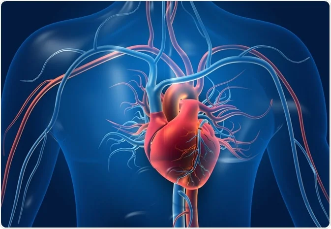
Overview of the Human Heart
The human heart is an amazing organ that is responsible for pumping blood throughout the body. It is made up of four chambers: the right atrium, the right ventricle, the left atrium, and the left ventricle. The heart also has four valves: the tricuspid valve, the pulmonary valve, the aortic valve, and the mitral valve.
These valves are open and close to control the flow of blood in and out of the heart. The heart is a muscle and it needs to be constantly pumping in order to circulate blood throughout the body. When you're resting, your heart rate should be about 60-80 beats per minute.
Anatomy of the Chambers of the Heart
The right atrium is the first chamber of the heart. It receives deoxygenated blood from the superior and inferior vena cava. The blood in the right atrium is dark red because it has not yet circulated through the body.
The left atrium is the third chamber of the heart. It receives oxygenated blood from the lungs. The blood in the left atrium is bright red because it has circulated through the body.
The left ventricle is the fourth and final chamber of the heart. It pumps oxygenated blood out to the body. The blood in the left ventricle is a darker red than the blood in the left atrium because it has not yet circulated through the body.
Functions of the Parts of the Heart
The heart is one of the most important organs in the human body, and it has a number of important functions. The right side of the heart pumps blood to the lungs, where it picks up oxygen. The left side of the heart then pumps the oxygenated blood throughout the rest of the body.
The heart also acts as a pump, circulating blood throughout the body. This helps to keep us healthy and functioning correctly. The heart is constantly working, even when we're asleep, and it can beat up to 150 times per minute!
Structures That Connect the Heart and Blood Vessels
Now, let’s move on to talking about the structures that connect the heart to the blood vessels. The superior vena cava and the inferior vena cava are two large veins responsible for carrying deoxygenated blood from different parts of your body back to the heart. This is what’s called systemic circulation, in which blood flows from a tissue or organ, through the veins, and then back again.
Next comes the pulmonary veins, which carry oxygenated blood from your lungs back to the left atrium of your heart. This is referred to as pulmonary circulation. Lastly, there’s an artery called the pulmonary trunk that carries deoxygenated blood from your heart to your lungs, where it can be re-oxygenated before it again flows back into your heart and throughout the rest of your body.
How Blood Flows Through the Heart
Once the heart's chambers are filled with blood, the heart starts to contract. As this happens, the right and left ventricles squeeze the blood into the pulmonary artery and aorta, respectively.
The pulmonary artery then carries oxygen-poor blood to the lungs to get replenished with oxygen. From there, it returns to the left atrium via the pulmonary veins. The aorta delivers oxygen-rich blood throughout your body.
This whole process is repeated again and again in a continuous loop. But how does this movement of blood keep happening?
As the right ventricle is squeezing out its contents, passive filling occurs in both atria due to pressure differences between them and their corresponding ventricles. This keeps new small amounts of blood entering both atria so that as soon as their corresponding ventricles have squeezed out their contents, they can start filling up again for more contractions and circulation.
Diseases Associated With the Human Heart
You may have heard of the various diseases associated with the human heart in the news, or from your medical professional. Cardiovascular diseases are among the most commonly diagnosed problems in patients throughout the world.
Most of these conditions, including heart attacks and strokes, are caused by a lack of blood supply to the heart, which can be due to a buildup of fatty deposits in an artery that leads to blockage. This can eventually cause an aneurysm or even a ruptured artery. Other conditions like cardiomyopathy, arrhythmia, and heart valve disease can also affect the normal functioning of the heart.
It is important to take preventive measures such as exercising regularly, maintaining healthy eating habits, and not smoking in order to reduce your susceptibility to these kinds of diseases and keep your heart strong and healthy.
valves and the characteristics and functions
You probably already know that the heart has four valves, but did you know why? These valves are in charge of keeping the blood flowing in one direction by regulating the pressure within the chambers of the heart. Let's take a closer look at them.
The tricuspid valve is located between the right atrium and right ventricle, while the mitral/bicuspid valve separates the left atrium and left ventricle. Both are made up of three flap-like structures called cusps that open and close to allow blood to flow through.
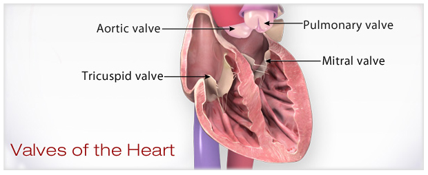
The pulmonary valve is located between the right ventricle and pulmonary artery while the aortic valve is located between the left ventricle and aorta, both made up of three cusps. The pulmonic and aortic valves close when pressure rises in either chamber so that no blood can return to it.
Valves ensure that oxygenated blood flows from your lungs to all parts of your body, while deoxygenated blood flows from all parts of your body back to your lungs for re-oxygenation. By regulating this flow of blood, they keep your heart functioning properly and keep you alive!
All in all, you now have a much better understanding of the heart and its function. You can see how the different chambers and valves work together to circulate blood throughout the body. You also have a clear view of the different layers of the heart and what each one does.
While we have covered a lot in this article, it's by no means an exhaustive look at the heart. There are still many things we don't know about this amazing organ. But, as research progresses, we are getting closer and closer to understanding all that the heart can do.


Perfect!
ReplyDelete